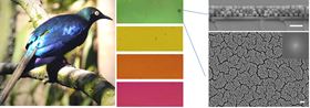
 (a) Photo of the African starling. Credit: Liliana D’Alba. (b) Optical image of thin films of SMNPs. Hue varies with the thickness and packing density of the particles. (c) Scanning electron micrographs of films made from nanoparticles of polydopamine seen in cross section.
Inspired by birds’ bright plumage, researchers from the University of California San Diego (UCSD), Scripps Institution of Oceanography, and The University of Akron have designed thin films of synthetic nanoparticles that mimic these colorful displays [Xiao et al., ACS Nano 9 (2015) 5454, http://dx.doi.org/10.1021/acsnano.5b01298].
Many bird species use arrays of self-assembled melanin nanoparticles in their feathers to create what is known as ‘structural color’ for mating display or camouflage (Fig. 1a). Structural color arises when spherical, rod-like, or disk-shaped melanin nanoparticles interact with light (Fig. 1b,c). Now researchers, led by Nathan C. Gianneschi at UCSD together with Matthew D. Shawkey and Ali Dhinojwala at Akron, have produced arrays of synthetic melanin nanoparticles (SMNPs) that create red, orange, yellow, and green colored films.
“We became interested in how melanin nanoparticles are packed in bird feathers to generate structural color,” explains Gianneschi, “so we began working to see if we could mimic that methodology to develop coloration that is not pigment based.”
The team synthesized melanin nanoparticles from polydopamine (PDA), the most common type of synthetic melanin, using a simple oxidative polymerization reaction of dopamine in water. The 146 ± 15 nm diameter nanoparticles are then dried on a surface to form self-assembled thin films.
The SMNPs have some very interesting properties, says Gianneschi, most notably a broad absorption spectrum and a high refractive index very similar to natural melanin. Scanning electron microscopy of green films reveals a thickness of 338 ± 9 nm with a color purity of 84%, while red films are 444 ± 15 nm thick, with 95% purity. The researchers note, however, that concentration changes during the evaporation process make it difficult to create uniform films at the centimeter scale.
This could be a turned into a potential advantage, says Gianneschi, as variations in film thickness could be used to create different colors. If the assembly process could be controlled, it might be possible to regulate the color variation. The researchers are now working on ways to improve control of the polymerization and self-assembly processes.
“In natural systems, this class of particle can be hollow, elliptical, rod-shaped etc.,” Gianneschi told Nano Today. “We aim to make all these shapes and then explore how they assemble to give other colors.”
SMNP films could have advantages over conventional colloidal arrays, say the researchers. Compared with polymeric particles, SMNPs generate more saturated colors and are less toxic, more biodegradable, and inherently biocompatible. This biomimetic approach to generating structural colors offers numerous opportunities for biocompatible photonic devices, believe the researchers.
This paper was originally published in Nano Today (2015), doi:10.1016/j.nantod.2015.06.009