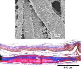
 CNT-decorated PCL nanofibers implanted in a rat calvarium defect could regenerate new bone in 8 weeks.
Biocompatible polymer scaffolds coated with a tangled mat of carbon nanotubes could provide a vital first foothold for regrowing cells, according to researchers from Dankook and Inha Universities in Korea [Patel et al., Acta Biomaterialia 108 (2020) 97-110, https://doi.org/10.1016/j.actbio.2020.03.012].
Scaffolds serve as support structures for growing cells to repair damaged or diseased tissue. But while synthetic biopolymers widely used in tissue engineering are biocompatible, bioactivity in terms of cell adhesion and growth is typically poor. To overcome this, surface structure or texture can be introduced to help cells stick, spread, and, in the case of stem cells, give the right cues to prompt their differentiation into specific cell types. Cleverly, Hae-Won Kim and his colleagues used the inherent dimensions of carbon nanotubes (CNTs) to create nanoscale topography on polymer nanofibers.
“We interfaced biopolymer nanofibers with CNTs in order to modulate multiple interactions of cells and tissues that are ultimately helpful for the tissue healing and bone regeneration process,” explains Kim.
The researchers used electrospinning to produce polycaprolactone (PCL) nanofibers with diameters of around 500 nm. After activating the nanofiber surfaces in alkaline solution to generate hydroxyl and carboxyl groups, the fibers are coated in a mat of CNTs with diameters of just 25 nm.
“We [explored] the issue that the bi-modal nanotopography generated by the nanofiber and nanotubes might be helpful for tissue repair process,” says Kim.
The bi-modal surface texture way appears to have a positive effect on a range of responses in a rat model system including inflammation, blood vessel growth (angiogenesis), and bone tissue regeneration. CNT-coated nanofibers show less inflammation when implanted, better angiogenic responses including new blood vessel formation, and accelerated bone regeneration with higher bone mineral density and elevated signs of osteogenesis.
“The unique nanotopographical features favor interactions with cells in terms of less inflammatory responses and promoted angiogenesis,” points out Kim, “which will ultimately help tissue healing and bone regeneration.”
Human bone marrow-derived mesenchymal stem cells (MSCs) seeded onto the CNT-coated nanofibers stick more readily to the surface and show accelerated differentiation into bone-forming osteogenic cells and increased mineralization and bone formation.
“This approach is very simple and can be applied to various polymeric surfaces,” adds Kim.
The researchers believe that it should also be possible to use the CNTs to load the scaffold surfaces with bioactive molecules such as drugs or growth factors, which could be released in the body to support specific healing processes.
“We now plan to examine more in vivo responses and apply the technique to different biopolymer surfaces such as scaffolds and spheres that are useful in the healing and regeneration of different tissues such as muscle and nerves,” adds Kim.