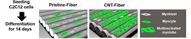
 Schematic showing myoblast cell differentiation on uncoated fibers and CNT-coated fibers over a period of 14 days.
A new carbon-based scaffold could help muscle tissue regenerate after injury or disease, according to researchers from University of Pittsburgh and Wright State University [Patel et al., Acta Biomaterialia (2016), DOI: 10.1016/j.actbio.2016.01.004].
Skeletal muscle has limited regenerative capabilities so the body needs extra assistance after trauma to help new tissue grow. One strategy is to use a scaffold material at the site of the damage to support the differentiation of progenitor cells (myoblasts) into myotubes as the first stage in muscle regeneration. Ultimately, myotubes form submicron-scale myofibrils, which in turn further organize themselves into fibers that bundle together to create functioning muscle. Any scaffolds aiming to promote tissue regeneration must mimic this hierarchical structure of micro- and nanoscale features.
Carbon is a potentially ideal scaffold material because it can be fabricated in various forms at different scales. Shilpa Sant of the University of Pittsburgh and Sharmila M. Mukhopadhyay of Wright State University made use of exactly this property in their hierarchical scaffolds.
“We chose carbon-based materials as a substrate for their good electrical conductivity, which is instrumental in promoting regeneration of electrically excitable skeletal muscle tissue,” explain Sant and Mukhopadhyay.
The team used conventional chemical vapor deposition (CVD) to grow a carpet of nanoscale carbon nanotubes (CNTs) on two different scaffold structures – microporous carbon foams and mats woven from carbon fibers.
Both hierarchical structures promote the adhesion, growth, and differentiation of progenitor cells into myocytes, with the CNT carpet appearing to provide a mechanical support for the growing cells until they become anchored, oriented, and differentiated into myotubes.
“Our materials demonstrate a greater regenerative potential as a result of synergetic effect of multi-scale structural and physicochemical features,” say Sant and Mukhopadhyay.
But the researchers found that only nanotube-coated fibrous mats stimulate the formation of aligned myotubes. The directional nature of the fibers in the mats appears to be essential in encouraging the fusion of myocytes into myotubes, the first step in functional muscle regeneration.
“The nanostructured CNT carpets offer fine control over nano-roughness and wettability facilitating myoblast adhesion, growth, and differentiation into myocytes,” explain the researchers. “Combined with the microscale aligned fibrous architecture of the carbon fabric substrate, this stimulates formation of multinucleated myotubes.”
Sant and Mukhopadhyay’s findings confirm that scaffolds for muscle regeneration must have an architecture with nanoscale features to encourage the differentiation of myoblasts into myocytes as well as microscale alignment cues to organize the fusion of myocytes into myotubes.
“These scaffolds could be used for regeneration of electrically excitable tissues such as skeletal muscles, neural tissues, and cardiac tissues, as well as for biosensors tailored to specific biomarkers or pathogens,” say the researchers.