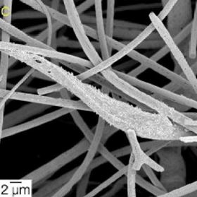
 SEM micrographs of MC3T3-E1 preosteoblasts cultured on 70S30C cotton-wool-like fibrous scaffolds.
A new cotton-wool-like bioactive glass scaffold material could make the repair of complex bone and tooth defects or injuries much easier.
Regeneration and repair strategies currently use synthetic, temporary scaffolds to support and promote the healing of bone and dental tissue. The ideal biocompatible scaffold material needs a three-dimensional structure that mimics the fibrous extracellular matrix (ECM) of bone. Bioactive glasses are a good option because they can form a rapid bond with bone and release silica and calcium ions to stimulate tissue repair.
Now a team of researchers from Imperial College London and the University of Manchester in the UK and Nagoya Institute of Technology in Japan have designed inorganic sol-gel solutions that can be electrospun into a cotton-wool-like three-dimensional bioactive glass scaffold [G. Poologasundarampillai, et al., Acta Biomaterialia 10 (2014) 3733–3746, DOI: 10.1016/j.actbio.2014.05.020].
“Bioactive glass can stimulate bone growth and bond with bone and soft tissue… [but] bulk glasses are brittle,” explains first author, Gowsihan Poologasundarampillai. “It is usually difficult to make into fibers without crystallization… [but] in this work we have produced flexible cotton-wool-like bioactive glass fibers.”
While the team employed a standard electrospinning technique, in which a jet of solution is forced out through a nozzle under an applied field, they did it under novel reaction and processing conditions to produce the flexible glass fibers without the need for a polymer binder. The sol-gel precursor solution, which is created by adding calcium nitrate tetrahydrate to a SiO2 solution, is key. The researchers believe that the presence of Ca2+ ions in the solution increase the charge density on the surface of the jet, splitting it into branches that form a mass of fine, individual fibers with diameters of 0.5-2 µm in a cotton-wool-like mass.
Once introduced into the body, the scaffold structure must allow the penetration and persistence of ECM-forming osteoblast cells to form new tissue.
“[The] structure is similar to the collagen fibrous structure of woven bone,” says Poologasundarampillai. “The large inter-fiber space in the cotton-wool-like material is ideal for colonization by cells, efficient diffusion of oxygen, and transport of nutrients into all parts of the scaffold.”
The researchers demonstrate that when the structure is immersed in simulated body fluid, a hydroxycarbonate apatite layer forms within 12 hours. Cells cultured on the scaffold material do not appear to experience any adverse effects and readily attach to the fibers and spread through the structure.
The new scaffold material could be particularly useful for dental implants, says Poologasundarampillai, because the flexible, cotton-wool-like material could be packed into awkward cavities or molded to fit complex defects. The team is also working on a variant containing antibacterial agents for wound dressing.
To download the article related to this news story, please click here.